Electrocardiogram (ECG): What It Is, What It Shows, How It’s Done & What It Diagnoses
Electrocardiogram (ECG): The Complete Guide
Understanding What It Is, What It Shows & How It Diagnoses Your Heart Health
❤️ Extended Summary: The Electrical Life of Your Heart
The electrocardiogram (ECG or EKG) is a cornerstone of modern cardiology, providing a painless and rapid window into your heart’s electrical activity. It captures the precise sequence of electrical impulses that trigger every heartbeat.
- Rapid Insights: Reveals heart rate, rhythm, and conduction speed in minutes.
- Critical Detection: Identifies signs of heart muscle damage, reduced blood flow (ischemia), and structural abnormalities.
- Emergency Tool: The first-line test for diagnosing heart attacks and life-threatening arrhythmias.
- Non-Invasive: Entirely painless with zero electrical risk to the body.
The electrocardiogram is one of the most widely used and important tests in modern medicine. Whether used during a routine check-up or in an emergency department, this quick diagnostic procedure records the electrical signals your heart produces with every beat. These impulses travel through the cardiac muscle in a precise sequence, which the ECG machine captures and displays as a predictable, wave-like pattern on a screen or paper.
The ECG Procedure: What to Expect
The process is straightforward and typically takes only 5 to 10 minutes from start to finish.
- Preparation: You lie flat on your back on an examination table, remaining as still and relaxed as possible.
- Lead Attachment: Small adhesive sensor patches (electrodes) are attached to your arms, legs, and across your chest.
- Recording: The machine reads your heart’s natural electricity for a few seconds to minutes.
- Review: The resulting trace is printed or displayed digitally for physician review.
Important Pre-Test Information
No fasting or special preparation is needed for a resting ECG. However, you should avoid applying lotions or creams to your chest, as these can interfere with the sensors. Most importantly, remain still; even deep breathing or minor muscle movement can affect the clarity of the recording.
Common Types of ECG Monitoring
| Type | Clinical Application |
|---|---|
| Resting ECG | The standard test for initial assessment while lying still. |
| 24-Hour Holter Monitor | A portable device used to capture intermittent arrhythmias over 24-48 hours. |
| Exercise Stress Test | Recorded during activity on a treadmill to detect exercise-induced ischemia. |
| Implantable Loop Recorder | A tiny device inserted for long-term monitoring (weeks to years). |
Decoding the Waveform: What an ECG Shows
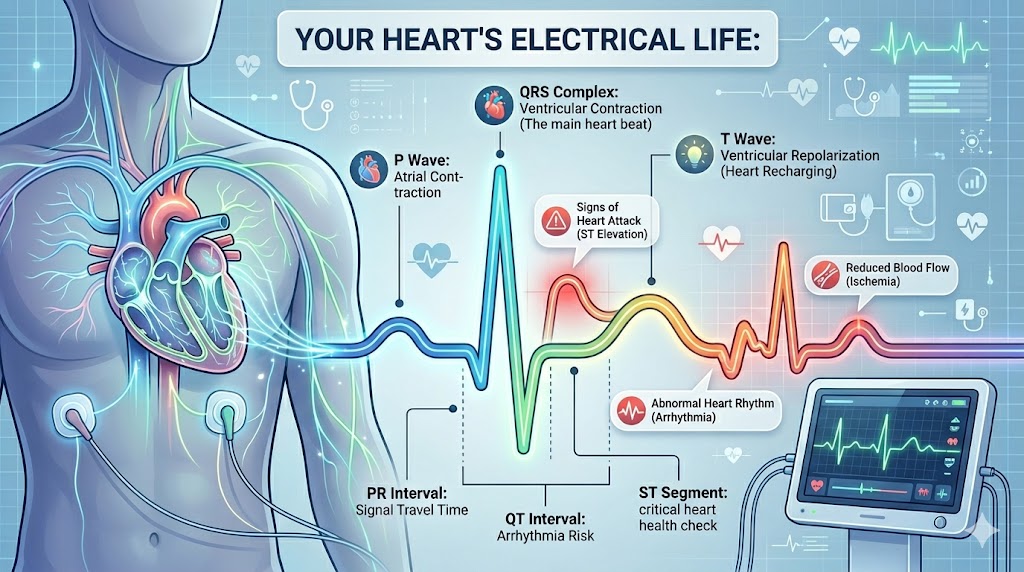
The lines on an ECG are not random; every peak and dip represents a specific electrical event in the heart.
- P Wave: Atrial contraction (upper chambers). Absence can suggest atrial fibrillation.
- QRS Complex: Ventricular contraction (main pumping chambers). Abnormalities here often indicate heart attack or blocks.
- T Wave: Ventricular “recharging” (repolarization). Changes may link to electrolyte imbalances or ischemia.
- ST Segment: Crucial for identifying active heart attacks (ST elevation or depression).
- PR/QT Intervals: Measures the timing of electrical travel; prolongations can indicate conduction delays or arrhythmia risks.
Clinical Diagnoses: Conditions Detected by ECG
ECG diagnosis covers a broad range of cardiac and systemic conditions:
- Heart Attack: Identifies ST elevation or pathological Q waves within minutes.
- Arrhythmias: Detects Atrial Fibrillation (AFib), Bradycardia (slow heart rate), and Tachycardia (fast heart rate).
- Structural Changes: Reveals signs of heart failure or chamber enlargement (hypertrophy).
- Electrolyte Imbalances: Abnormal levels of potassium, calcium, or magnesium leave identifiable “fingerprints”.
- Genetic Syndromes: Identifies conditions like Wolff-Parkinson-White, Brugada, and Long QT syndrome.
- Inflammation: Distinguishes Pericarditis (sac inflammation) from a heart attack.
Reliability and Next Steps
While highly reliable, a normal resting ECG does not entirely rule out heart disease. Significant coronary artery disease may show no abnormalities at rest, and intermittent arrhythmias may not occur during the short recording window. To get a complete picture, clinicians often combine ECGs with blood tests (troponins), echocardiograms (ultrasound), or CT angiography.
Frequently Asked Questions
Is an ECG painful? No. You will only feel light pressure from the adhesive electrodes.
Do I need to fast? No fasting is required for a standard resting ECG.
ECG vs. Echocardiogram? An ECG records electrical activity, while an echocardiogram uses ultrasound to see physical structure and pumping function.
Don’t Ignore Your Heart’s Signals
If you are experiencing chest pain, palpitations, or unexplained breathlessness, early detection is vital.
Last Updated: May 2026 | Reviewed in collaboration with specialists.
⚕️ MEDICAL DISCLAIMER
This article is for informational purposes only and does not constitute medical advice. Always consult a qualified healthcare professional regarding any symptoms or concerns about your heart health.
Read Also