Ultrasound technology: a cornerstone of modern medicine for decades, providing a non-invasive, safe, and effective way to see inside the human body. Whether you’ve seen one during a pregnancy check-up or for a medical diagnosis, you may have wondered: What is an ultrasound, and how does it actually work?
At its core, an ultrasound uses high-frequency sound waves—far above the range of human hearing—to create images of internal organs, tissues, and blood vessels. Unlike X-rays, which use ionizing radiation, ultrasound is completely radiation-free, making it a preferred choice for many medical applications, especially during pregnancy.
The Science Behind the Sound Waves
Think of it like a bat using echolocation. A special device called a transducer acts as both a speaker and a microphone. It sends out short pulses of inaudible sound waves into the body. When these waves encounter different tissues (like muscle, fat, bone, or fluid), they bounce back, or “echo.”
The transducer then listens for these echoes. The time it takes for an echo to return, and the strength of the returning signal, provides crucial information.
- Time: The longer it takes for an echo to return, the deeper the structure is.
- Strength: The strength of the echo tells us about the type of tissue. For example, a dense structure like bone will produce a strong echo, while a fluid-filled cavity will produce a weak one.
A powerful computer processes all this information in real-time, creating a detailed, two-dimensional image on a screen. This image, called a sonogram, allows doctors to visualize internal structures without needing to make a single incision.
Understanding Different Ultrasound Modes
Modern ultrasound machines are incredibly versatile, offering different imaging modes to suit specific diagnostic needs.
- B-mode (Brightness mode): This is the most common mode, producing the familiar 2D black-and-white image. The brightness of each pixel on the screen corresponds to the strength of the returning echo, creating a cross-sectional view of the anatomy. This is used for general organ and fetal imaging.
- M-mode (Motion mode): This mode is used to visualize moving structures. It captures a single scan line over time, showing how a structure (like a heart valve) moves. M-mode is essential in echocardiography to assess heart function and valve movement.
- Doppler Ultrasound: This specialized mode uses the Doppler effect to measure the direction and speed of blood flow. By analyzing changes in the frequency of the returning sound waves, a color-coded image or an audible signal is created to show blood flow. This is critical for detecting blood clots, blockages, or other vascular issues.
- 3D and 4D Ultrasound: These advanced techniques create volumetric images. A 3D ultrasound acquires multiple 2D images and reconstructs them into a static three-dimensional view. A 4D ultrasound takes it a step further by capturing 3D images in real-time, creating a live video of the fetus or an internal organ, often providing invaluable insight for diagnosis and parent-to-be bonding.
Diverse Applications Across Medicine
The versatility of ultrasound makes it indispensable across various medical fields.
- Obstetrics and Gynecology: This is perhaps the most well-known use. Ultrasounds are used to monitor fetal development, check the baby’s position and growth, and determine the due date. The detail offered by 3D and 4D imaging can reveal anatomical abnormalities that are difficult to see on a standard 2D scan.
- Cardiology: Echocardiograms are a type of ultrasound used to examine the heart. They can assess heart function, blood flow, and detect abnormalities like congenital heart defects.
- Vascular Medicine: Doppler ultrasound is a key tool for diagnosing conditions like Deep Vein Thrombosis (DVT), arterial blockages, and aneurysms. It provides a non-invasive way to visualize blood vessels and blood flow patterns.
- Abdominal Imaging: Doctors use ultrasounds to examine organs like the liver, kidneys, gallbladder, and pancreas to diagnose conditions such as gallstones or kidney stones.
- Musculoskeletal Imaging: It can visualize muscles, tendons, ligaments, and joints to diagnose sprains, tears, and other injuries.
- Emergency Medicine: In emergency rooms, portable ultrasound devices are crucial for quick assessments of internal bleeding or fluid build-up, helping doctors make rapid, life-saving decisions.
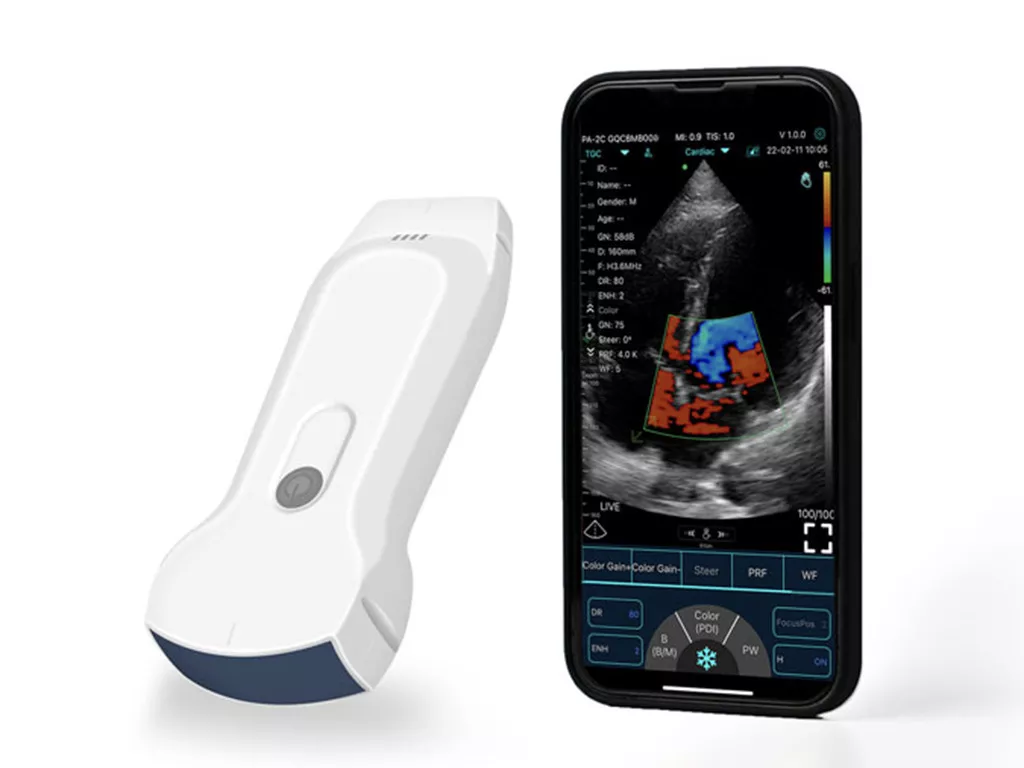
The Rise of Portable Ultrasound Devices
Traditionally, ultrasound machines were large, stationary pieces of equipment found only in hospitals and clinics. However, recent technological advancements have led to the development of portable ultrasound devices. These compact, handheld units have revolutionized healthcare by bringing diagnostic imaging directly to the patient’s bedside, a remote clinic, or even a home visit.
This portability offers significant benefits:
- Increased Accessibility: It makes diagnostic imaging available in rural or underserved areas where large hospital equipment isn’t feasible.
- Rapid Diagnosis: Emergency responders and doctors can quickly assess a patient’s condition on the spot, leading to faster and more effective treatment.
- Enhanced Patient Care: Instead of having to transport a patient to a separate department for a scan, a doctor can perform the ultrasound right at the bedside.
A prime example of this innovation is the Konted C10tx, a state-of-the-art portable ultrasound device. It’s designed for ease of use and delivers high-quality imaging in a compact, wireless form factor. The Konted C10tx enables healthcare professionals to provide on-demand diagnostic services, bridging the gap between traditional medicine and modern, mobile healthcare. Its user-friendly interface and superior image clarity make it an invaluable tool for a wide range of medical applications, from routine check-ups to critical care.
The Future of Ultrasound: AI and Beyond
The future of ultrasound technology is as exciting as its past. Artificial intelligence (AI) is already being integrated into devices to assist with image acquisition and interpretation, making the technology even more accessible to non-specialists. AI algorithms can help automatically identify organs, measure structures, and even flag potential abnormalities, reducing human error and saving time.
Furthermore, the rise of telemedicine and remote patient monitoring means that portable ultrasound devices, like the Konted C10tx, will play a central role. A doctor could guide a patient or a less-experienced healthcare provider through a scan remotely, allowing for expert diagnosis regardless of physical location. This fusion of powerful technology and mobile connectivity is set to make high-quality diagnostic imaging a reality for everyone, everywhere.
Conclusion: A Window into Your Body’s Health
In conclusion, ultrasound is a powerful and safe diagnostic tool that uses sound waves to create a window into the human body. From monitoring new life to guiding emergency procedures and pioneering remote medicine, its applications are vast and continue to expand with the evolution of technology. It remains a key pillar of modern medicine, providing essential insights without the need for invasive procedures.
Sources
- American Institute of Ultrasound in Medicine (AIUM)
- Radiological Society of North America (RSNA)
- National Institute of Biomedical Imaging and Bioengineering (NIBIB)
⚕️ Important Disclaimer
This article is intended for informational purposes only. It does not replace professional medical advice, diagnosis, or treatment. Always consult with qualified healthcare professionals regarding your health. Never alter your treatment plan without medical guidance.
In the event of an emergency, contact local emergency services or visit the nearest hospital immediately. This website and its authors are not liable for outcomes resulting from reliance on the content herein. Readers must take full responsibility for their medical decisions.